How to Take 3D Pictures Inside Human Tissue Using Near-Infrared Light
A system that uses flexible fiber optic cables to map the inside of complex body parts by shining light through them and measuring how it scatters.
Original patent title: “USRE38800E1 - NIR clinical opti-scan system”
A system that uses flexible fiber optic cables to map the inside of complex body parts by shining light through them and measuring how it scatters. Granted to Research Foundation of the State University of New York in 2005 with 42 claims and 25 forward citations.
Coverage
What does this patent actually cover?
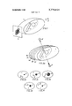
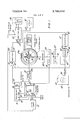
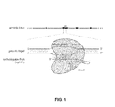
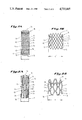
This patent describes a way to create 3D images of internal body structures by shining near-infrared light into tissue and collecting the light that bounces back out. It uses an adjustable support structure, like a flexible frame, that holds a large array of fiber optic cables against the uneven surface of a body part. These fibers act as both the light source and the light collector, allowing the system to capture data from many different points simultaneously. A detector then measures this scattered light, and a computer uses that data to calculate what the internal tissue looks like in three dimensions.
The gap
What does this patent NOT cover?
- Does not cover imaging techniques that rely on X-rays or ionizing radiation.
- Does not cover systems that lack the specific adjustable fiber-holding assembly for irregular surfaces.
- Does not cover ultrasound or MRI-based imaging methods.
- Does not cover imaging that requires invasive surgical entry into the body.
These exclusions are unique to PatentBrief — derived from the actual claim language, not patent-office boilerplate.
Key facts
What made this novel
The innovation lies in the adjustable fiber array that physically conforms to the non-uniform, curved surface of a body part, ensuring consistent contact and data collection across complex shapes.
Schematic visualization of the patent's claim structure. Hand-drawn diagrams in progress for each landmark patent.
Where you've seen this
Real-world examples
Optical breast imaging systems
Near-infrared diffuse optical tomography (DOT) scanners
Non-invasive tissue diagnostic devices
Why it matters
The bigger picture
This technology is significant because it provides a non-invasive way to look inside soft tissue, which is often difficult to image clearly with traditional methods. By using near-infrared light, it can detect differences in how light is absorbed or scattered by different types of tissue, potentially helping to identify tumors or other abnormalities without exposing the patient to harmful radiation.
Filed
June 27, 2002
Granted
September 20, 2005
Market context
Who's building on this
Companies in this space
Research institutions and medical device companies focused on diffuse optical tomography (DOT) continue to refine these concepts. The original assigneeassigneeThe entity that owns the patent — usually the inventor's employer or a company.Read more →, the Research Foundation of the State University of New York, has been a key player in advancing this specific approach to optical imaging.
Market impact
This patent helped formalize the hardware requirements for practical optical tomography, moving it from a theoretical physics concept toward a deployable medical diagnostic tool. It established a framework for using flexible fiber arrays to solve the problem of imaging irregular biological surfaces, influencing subsequent designs in non-invasive diagnostic equipment.
Claim 1 — Plain English
What this patent covers
This patent describes a way to create 3D images of internal body structures by shining near-infrared light into tissue and collecting the light that bounces back out. It uses an adjustable support structure, like a flexible frame, that holds a large array of fiber optic cables against the uneven surface of a body part. These fibers act as both the light source and the light collector, allowing the system to capture data from many different points simultaneously. A detector then measures this scattered light, and a computer uses that data to calculate what the internal tissue looks like in three dimensions.
The clever bit
The innovation lies in the adjustable fiber array that physically conforms to the non-uniform, curved surface of a body part, ensuring consistent contact and data collection across complex shapes.
What it does not cover
- Does not cover imaging techniques that rely on X-rays or ionizing radiation.
- Does not cover systems that lack the specific adjustable fiber-holding assembly for irregular surfaces.
- Does not cover ultrasound or MRI-based imaging methods.
- Does not cover imaging that requires invasive surgical entry into the body.
Patent timeline
Application submitted to the patent office
Application published, typically 18 months after filing
Patent officially issued
PatentBrief Score
Impact Score
Strong
Citation count
28/40
Moderately cited
Claim breadth
20/20
Very broad protection
Recency
0/20
Older than 20 years
Assignee scale
20/20
Major company or institution
PatentBrief Impact Score — based on citation count, claim breadth, recency, and assignee scale. Not a legal assessment.
Heuristic Value Estimate
What this patent might be worth
$32K – $104K
Midpoint $65K · expired or expiring · industry ×1.5
Heuristic only — blends forward/backward citation counts, claim scope, time remaining, litigation history, and CPC-derived industry baseline. Real valuations need a professional appraisal.
Patent Claims
0 independent claims · 1 dependent
Claims are the legal boundaries of the patent. An independent claim stands alone. A dependent claim adds limitations to its parent, narrowing — but not broadening — the scope.
The original legal language
Original claims
42 claims as filed with the patent office.
Concepts involved
Citations
Patent lineage
Cite this patent
Barbour, R. L. (2005). How to Take 3D Pictures Inside Human Tissue Using Near-Infrared Light (U.S. Patent No. RE38,800). U.S. Patent and Trademark Office. https://patentbrief.org/patent/us/RE38800/digital-video-recorder-dvr
Auto-generated from the patent record. Double-check author order and the issue date against the official USPTO document before submitting.
Embed
Add this patent to your site
Drop this plain-English patent card into any blog post or article — free, no signup. It always links back to the full breakdown here.
<div data-patentlens-widget data-patent-number="USRE38800"></div>
<script src="https://patentbrief.org/embed.js" async></script>
Stay in the loop
Get a weekly digest of new patents.
One email per week. No spam. Unsubscribe anytime.
Keep exploring
Related patents you should know
US 4683195 · 1987
How to Make Billions of Copies of a DNA Segment
This patent describes the Polymerase Chain Reaction (PCR), a method to rapidly create many copies of a specific piece of DNA or RNA, enabling its detection and analysis.
Cetus Corp
US 8697359 · 2014
How to Edit Genes in Human Cells Using an Engineered CRISPR System
This patent describes an engineered CRISPR-Cas9 system for precisely cutting DNA in eukaryotic cells to change how genes work, opening the door for gene editing in complex organisms.
Massachusetts Institute of Technology
US 7657849 · 2010
How the iPhone's Slide-to-Unlock Gesture Works
Apple's 2010 patent describes unlocking a device by dragging a specific graphical image across the touchscreen along a predefined path, a gesture that became iconic with the original iPhone.
Apple Inc
US 4733665 · 1988
How Doctors Implant a Permanent Stent Using a Balloon
This patent describes the method for placing a permanent, expandable wire mesh tube inside a blood vessel or other body tube using a balloon-tipped catheter to widen it and keep it open.
Expandable Grafts Partnership
US 4965188 · 1990
How to Make Many Copies of a DNA Piece with Heat
This patent describes the Polymerase Chain Reaction (PCR) method, a technique to make millions of copies of a specific DNA segment using a heat-resistant enzyme and repeated temperature changes.
Cetus Corp
US 4235871 · 1980
How to Encapsulate Active Materials in Lipid Bubbles Efficiently
This patent describes a method for trapping biologically active substances inside tiny, multi-layered fat bubbles called liposomes, using a specific water-in-oil emulsion and gel-forming process to improve how much material gets captured.
Individual
Semantically similar
You might also find these interesting
US 3778614 · 1973 · EMI Ltd
How Early CT Scans Created Detailed Body Images
US 9677869 · 2017 · Perimeter Medical Imaging Inc
How to Create Wide-Field Images of Tissue Using OCT Scanners
US 3789832 · 1974
Using Nuclear Magnetic Resonance to Detect Cancer in Tissue
US RE50510 · 2025 · Sony Group Corp
How to Embed Tiny Optical Components Directly into Circuit Boards
More to explore
More in Biotech & Medicine
US 4683195 · 1987 · Cetus Corp
How to Make Billions of Copies of a DNA Segment
US 8697359 · 2014 · Massachusetts Institute of Technology
How to Edit Genes in Human Cells Using an Engineered CRISPR System
US 4733665 · 1988 · Expandable Grafts Partnership
How Doctors Implant a Permanent Stent Using a Balloon
US 4965188 · 1990 · Cetus Corp
How to Make Many Copies of a DNA Piece with Heat
New to patents?
Common Questions
Frequently Asked Questions
What does How to Take 3D Pictures Inside Human Tissue Using Near-Infrared Light cover?
A system that uses flexible fiber optic cables to map the inside of complex body parts by shining light through them and measuring how it scatters.
Who owns patent US RE38800?
Research Foundation of the State University of New York owns this patent, granted in 2005.
When does this patent expire?
This patent has expired and is now in the public domain — anyone can use the invention freely.
What is patent US RE38800 cited by?
This patent has been cited by 25 later patents that build on its ideas.
What problem does this patent solve?
This technology is significant because it provides a non-invasive way to look inside soft tissue, which is often difficult to image clearly with traditional methods. By using near-infrared light, it can detect differences in how light is absorbed or scattered by different types of tissue, potentially helping to identify tumors or other abnormalities without exposing the patient to harmful radiation.
What does this patent NOT cover?
Does not cover imaging techniques that rely on X-rays or ionizing radiation.
Patent monitoring