How Early CT Scans Created Detailed Body Images
This 1973 patent describes a method for using X-rays from many angles to build a detailed 2D image of the inside of a body, like a slice of a CT scan.
Original patent title: “Method and apparatus for measuring x- or {65 -radiation absorption or transmission at plural angles and analyzing the data”
This 1973 patent describes a method for using X-rays from many angles to build a detailed 2D image of the inside of a body, like a slice of a CT scan. Granted to EMI Ltd in 1973 with 37 claims and 163 forward citations, and it is now in the public domain.
Coverage
What does this patent actually cover?
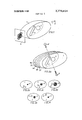
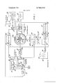
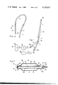
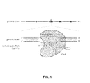
This patent details a method for creating a 2D image of a body slice using penetrating radiation, like X-rays. Radiation is sent through the body from many different angles, passing through many paths. Each path's 'transmission' (how much radiation gets through) is measured. The patent claimsclaimsThe numbered statements at the end of a patent that legally define what the inventor owns.Read more → a way to use these measurements, especially by 'successive approximations' where calculations are repeatedly refined, to figure out the absorption or transmission of tiny, individual elements within the body's 2D matrix. These refined values are then used to create a visual representation, like on a screen or photo, showing the internal structure. For example, imagine shining X-rays through a slice of your arm from the front, then the side, then diagonally, and using those readings to map out the density of bone versus muscle in each tiny spot.
The gap
What does this patent NOT cover?
- Methods that only use radiation from a single angle.
- Imaging techniques that don't involve measuring radiation transmission or absorption.
- Creating 3D images instead of 2D slices.
- Methods that don't use a 'successive approximation' process to calculate internal element values.
- Using radiation sources other than X-rays or gamma rays.
These exclusions are unique to PatentBrief — derived from the actual claim language, not patent-office boilerplate.
Key facts
What made this novel
The core innovation was developing a mathematical method, specifically 'successive approximations,' to reconstruct a detailed 2D image from numerous, incomplete X-ray measurements taken at different angles. This allowed for the differentiation of absorption coefficients of neighboring elements, which was crucial for creating diagnostic images.
The Patent Drawing
Schematic visualization of the patent's claim structure. Hand-drawn diagrams in progress for each landmark patent.
Where you've seen this
Real-world examples
Early CT scanners
The technology behind modern CT imaging
Why it matters
The bigger picture
This patent is foundational to the development of computed tomography (CT) scanning. Sir Godfrey Hounsfield, one of the inventors, won the Nobel Prize in Physiology or Medicine in 1979 for his work on this technology. CT scans revolutionized medical diagnostics by providing detailed cross-sectional images of the body, enabling doctors to see internal structures with unprecedented clarity.
Filed
December 27, 1971
Granted
December 11, 1973
Market context
Who's building on this
Companies in this space
The foundational principles of this patent are embedded in virtually all modern CT scanner manufacturers, including GE Healthcare, Siemens Healthineers, Philips, and Canon Medical Systems. Research continues to refine image reconstruction algorithms and detector technologies.
Market impact
This patent enabled the creation of the first practical CT scanners, fundamentally changing medical imaging. It established a new category of diagnostic tools, leading to widespread adoption in hospitals worldwide and significantly improving the diagnosis of a vast range of medical conditions.
Claim 1 — Plain English
What this patent covers
This patent details a method for creating a 2D image of a body slice using penetrating radiation, like X-rays. Radiation is sent through the body from many different angles, passing through many paths. Each path's 'transmission' (how much radiation gets through) is measured. The patent claims a way to use these measurements, especially by 'successive approximations' where calculations are repeatedly refined, to figure out the absorption or transmission of tiny, individual elements within the body's 2D matrix. These refined values are then used to create a visual representation, like on a screen or photo, showing the internal structure. For example, imagine shining X-rays through a slice of your arm from the front, then the side, then diagonally, and using those readings to map out the density of bone versus muscle in each tiny spot.
The clever bit
The core innovation was developing a mathematical method, specifically 'successive approximations,' to reconstruct a detailed 2D image from numerous, incomplete X-ray measurements taken at different angles. This allowed for the differentiation of absorption coefficients of neighboring elements, which was crucial for creating diagnostic images.
What it does not cover
- Methods that only use radiation from a single angle.
- Imaging techniques that don't involve measuring radiation transmission or absorption.
- Creating 3D images instead of 2D slices.
- Methods that don't use a 'successive approximation' process to calculate internal element values.
- Using radiation sources other than X-rays or gamma rays.
Patent timeline
Application submitted to the patent office
Application published, typically 18 months after filing
Patent officially issued
Patent enters public domain
This patent is in the public domain
See the Freedom to Build guide — what is free to use, what is not, and how to cite this patent.
PatentBrief Score
Impact Score
Strong
Citation count
40/40
Highly cited
Claim breadth
20/20
Very broad protection
Recency
0/20
Older than 20 years
Assignee scale
0/20
Independent or smaller assigneeassigneeThe entity that owns the patent — usually the inventor's employer or a company.Read more →
PatentBrief Impact Score — based on citation count, claim breadth, recency, and assignee scale. Not a legal assessment.
Heuristic Value Estimate
What this patent might be worth
$158K – $507K
Midpoint $317K · expired or expiring · industry ×2.2
Heuristic only — blends forward/backward citation counts, claim scope, time remaining, litigation history, and CPC-derived industry baseline. Real valuations need a professional appraisal.
Patent Claims
0 independent claims · 1 dependent
Claims are the legal boundaries of the patent. An independent claim stands alone. A dependent claim adds limitations to its parent, narrowing — but not broadening — the scope.
The original legal language
Original claims
37 claims as filed with the patent office.
Concepts involved
Citations
Patent lineage
Cite this patent
Newbold, H. G. (1973). How Early CT Scans Created Detailed Body Images (U.S. Patent No. 3,778,614). U.S. Patent and Trademark Office. https://patentbrief.org/patent/us/3778614/ct-scanner-hounsfield
Auto-generated from the patent record. Double-check author order and the issue date against the official USPTO document before submitting.
Embed
Add this patent to your site
Drop this plain-English patent card into any blog post or article — free, no signup. It always links back to the full breakdown here.
<div data-patentlens-widget data-patent-number="US3778614"></div>
<script src="https://patentbrief.org/embed.js" async></script>
Stay in the loop
Get a weekly digest of new patents.
One email per week. No spam. Unsubscribe anytime.
Keep exploring
Related patents you should know
US 4683195 · 1987
How to Make Billions of Copies of a DNA Segment
This patent describes the Polymerase Chain Reaction (PCR), a method to rapidly create many copies of a specific piece of DNA or RNA, enabling its detection and analysis.
Cetus Corp
US 8697359 · 2014
How to Edit Genes in Human Cells Using an Engineered CRISPR System
This patent describes an engineered CRISPR-Cas9 system for precisely cutting DNA in eukaryotic cells to change how genes work, opening the door for gene editing in complex organisms.
Massachusetts Institute of Technology
US 7657849 · 2010
How the iPhone's Slide-to-Unlock Gesture Works
Apple's 2010 patent describes unlocking a device by dragging a specific graphical image across the touchscreen along a predefined path, a gesture that became iconic with the original iPhone.
Apple Inc
US 4733665 · 1988
How Doctors Implant a Permanent Stent Using a Balloon
This patent describes the method for placing a permanent, expandable wire mesh tube inside a blood vessel or other body tube using a balloon-tipped catheter to widen it and keep it open.
Expandable Grafts Partnership
US 4965188 · 1990
How to Make Many Copies of a DNA Piece with Heat
This patent describes the Polymerase Chain Reaction (PCR) method, a technique to make millions of copies of a specific DNA segment using a heat-resistant enzyme and repeated temperature changes.
Cetus Corp
US 4235871 · 1980
How to Encapsulate Active Materials in Lipid Bubbles Efficiently
This patent describes a method for trapping biologically active substances inside tiny, multi-layered fat bubbles called liposomes, using a specific water-in-oil emulsion and gel-forming process to improve how much material gets captured.
Individual
Semantically similar
You might also find these interesting
US RE38800 · 2005 · Research Foundation of the State University of New York
How to Take 3D Pictures Inside Human Tissue Using Near-Infrared Light
US 3789832 · 1974
Using Nuclear Magnetic Resonance to Detect Cancer in Tissue
US 3927193 · 1975 · F Hoffmann La Roche AG
Using Radioactive Antibodies to Find Tumors Inside the Body
US 4195637 · 1980 · Schneider Medintag AG
Catheter System for Opening and Closing Body Passages
More to explore
More in Biotech & Medicine
US 4683195 · 1987 · Cetus Corp
How to Make Billions of Copies of a DNA Segment
US 8697359 · 2014 · Massachusetts Institute of Technology
How to Edit Genes in Human Cells Using an Engineered CRISPR System
US 4733665 · 1988 · Expandable Grafts Partnership
How Doctors Implant a Permanent Stent Using a Balloon
US 4965188 · 1990 · Cetus Corp
How to Make Many Copies of a DNA Piece with Heat
New to patents?
Common Questions
Frequently Asked Questions
What does How Early CT Scans Created Detailed Body Images cover?
This 1973 patent describes a method for using X-rays from many angles to build a detailed 2D image of the inside of a body, like a slice of a CT scan.
Who owns patent US 3778614?
EMI Ltd owns this patent, granted in 1973.
When does this patent expire?
This patent has expired and is now in the public domain — anyone can use the invention freely.
What is patent US 3778614 cited by?
This patent has been cited by 163 later patents that build on its ideas.
What problem does this patent solve?
This patent is foundational to the development of computed tomography (CT) scanning. Sir Godfrey Hounsfield, one of the inventors, won the Nobel Prize in Physiology or Medicine in 1979 for his work on this technology. CT scans revolutionized medical diagnostics by providing detailed cross-sectional images of the body, enabling doctors to see internal structures with unprecedented clarity.
What does this patent NOT cover?
Methods that only use radiation from a single angle.
Patent monitoring