How Doctors Use CT Scans and X-rays to Check Heart Arteries
This patent describes a method for precisely evaluating blockages in heart arteries by combining a single X-ray image with a detailed 3D CT scan, then calculating blood flow.
Original patent title: “Systems and methods for registration of angiographic projections with computed tomographic data”
This patent describes a method for precisely evaluating blockages in heart arteries by combining a single X-ray image with a detailed 3D CT scan, then calculating blood flow. Granted to GE Precision Healthcare in 2024 with 19 claims and 6 forward citations, and it is expected to expire in 2040.
Coverage
What does this patent actually cover?
The patent outlines a way to image heart arteries using two types of scans. First, it acquires detailed 3D computed tomography (CT) imaging data depicting the patient's entire coronary tree (ClaimclaimA numbered sentence at the end of a patent that legally defines what the inventor owns. The most important section.Read more → 1). After the CT scan, it acquires a single 2D X-ray image, called an angiographic projection, of a specific coronary artery, selecting the view with the least amount of vessel superimposition (Claim 1). The system then "registers" or aligns this single angiographic projection to the 3D CT imaging data. This registration involves segmenting the central paths, or "centerlines," of the coronary tree from the CT data, determining their length and three-dimensional shape, and then using this information to align the 2D X-ray (Claim 1). Finally, it determines the Fractional Flow Reserve (FFR) of the coronary artery from this registered data, which indicates how well blood is flowing through it (Claim 1). For example, if a patient has a narrowed artery, this method could help doctors assess if the blockage is severe enough to require a stent (Claim 4).
The gap
What does this patent NOT cover?
- Does not cover methods that require multiple angiographic projections for 3D reconstruction of the coronary artery.
- Does not cover registration methods that do not rely on segmenting and matching the 3D shape and length of coronary artery centerlines.
- Does not cover using other 3D imaging modalities, like MRI, instead of computed tomography (CT) data for the initial 3D model.
- Does not cover diagnostic methods for vessels other than coronary arteries or metrics other than Fractional Flow Reserve (FFR).
- Does not cover systems that acquire the single angiographic projection without selecting for the least amount of vessel superimposition.
These exclusions are unique to PatentBrief — derived from the actual claim language, not patent-office boilerplate.
Key facts
What made this novel
The clever part is efficiently combining a detailed 3D CT scan with just a single 2D X-ray angiogram. It uses the precise 3D centerline information from the CT scan to accurately align the 2D X-ray, allowing for a reliable calculation of blood flow (FFR) with minimal additional imaging.
The Patent Drawing
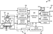
Schematic visualization of the patent's claim structure. Hand-drawn diagrams in progress for each landmark patent.
Where you've seen this
Real-world examples
Cardiac catheterization labs
Interventional cardiology suites
Advanced medical imaging systems for heart diagnostics
Software for FFR calculation from hybrid imaging data
Why it matters
The bigger picture
This technology matters because it offers a less invasive and potentially faster way to diagnose coronary artery disease. By combining existing 3D CT data with a single 2D angiogram, it can reduce the amount of radiation and contrast dye a patient receives compared to traditional methods that might require multiple angiograms. This helps doctors make more informed decisions about treatments like stent placement, improving patient care and outcomes.
Filed
March 10, 2020
Granted
July 30, 2024
Market context
Who's building on this
Companies in this space
GE Precision Healthcare, the assigneeassigneeThe entity that owns the patent — usually the inventor's employer or a company.Read more →, is a major player in medical imaging and continues to develop advanced diagnostic tools for cardiology. Other large medical technology companies like Siemens Healthineers, Philips Healthcare, and Canon Medical Systems also invest heavily in integrated imaging solutions for cardiac assessment, often exploring ways to combine different imaging modalities for comprehensive diagnosis.
Market impact
This patent contributes to the ongoing shift towards less invasive and more precise cardiac diagnostics. By streamlining the FFR calculation process with fewer angiographic projections, it could lead to reduced procedure times, lower patient exposure to radiation and contrast agents, and more efficient use of resources in interventional cardiology. This could make FFR assessment more widely accessible and integrated into routine diagnostic workflows, potentially improving patient outcomes for coronary artery disease.
Claim 1 — Plain English
What this patent covers
The patent outlines a way to image heart arteries using two types of scans. First, it acquires detailed 3D computed tomography (CT) imaging data depicting the patient's entire coronary tree (Claim 1). After the CT scan, it acquires a single 2D X-ray image, called an angiographic projection, of a specific coronary artery, selecting the view with the least amount of vessel superimposition (Claim 1). The system then "registers" or aligns this single angiographic projection to the 3D CT imaging data. This registration involves segmenting the central paths, or "centerlines," of the coronary tree from the CT data, determining their length and three-dimensional shape, and then using this information to align the 2D X-ray (Claim 1). Finally, it determines the Fractional Flow Reserve (FFR) of the coronary artery from this registered data, which indicates how well blood is flowing through it (Claim 1). For example, if a patient has a narrowed artery, this method could help doctors assess if the blockage is severe enough to require a stent (Claim 4).
The clever bit
The clever part is efficiently combining a detailed 3D CT scan with just a single 2D X-ray angiogram. It uses the precise 3D centerline information from the CT scan to accurately align the 2D X-ray, allowing for a reliable calculation of blood flow (FFR) with minimal additional imaging.
What it does not cover
- Does not cover methods that require multiple angiographic projections for 3D reconstruction of the coronary artery.
- Does not cover registration methods that do not rely on segmenting and matching the 3D shape and length of coronary artery centerlines.
- Does not cover using other 3D imaging modalities, like MRI, instead of computed tomography (CT) data for the initial 3D model.
- Does not cover diagnostic methods for vessels other than coronary arteries or metrics other than Fractional Flow Reserve (FFR).
- Does not cover systems that acquire the single angiographic projection without selecting for the least amount of vessel superimposition.
Patent timeline
Application submitted to the patent office
Application published, typically 18 months after filing
Patent officially issued
Patent enters public domain
PatentBrief Score
Impact Score
Strong
Citation count
17/40
Early citations
Claim breadth
13/20
Broad claimsclaimsThe numbered statements at the end of a patent that legally define what the inventor owns.Read more →
Recency
20/20
Granted within 5 years
Assignee scale
20/20
Major company or institution
PatentBrief Impact Score — based on citation count, claim breadth, recency, and assignee scale. Not a legal assessment.
Heuristic Value Estimate
What this patent might be worth
$206K – $659K
Midpoint $412K · 13.5 yr remaining · industry ×2.2
Heuristic only — blends forward/backward citation counts, claim scope, time remaining, litigation history, and CPC-derived industry baseline. Real valuations need a professional appraisal.
Claim text not yet imported for this patent
The original legal language
Original claims
19 claims as filed with the patent office.
Concepts involved
Citations
Patent lineage
Cite this patent
Ramus, L., Amelot, S., & Vaillant, R. (2024). How Doctors Use CT Scans and X-rays to Check Heart Arteries (U.S. Patent No. 12,048,575). U.S. Patent and Trademark Office. https://patentbrief.org/patent/us/12048575/systems-and-methods-for-registration-of-angiographic-projections-with-computed-t
Auto-generated from the patent record. Double-check author order and the issue date against the official USPTO document before submitting.
Embed
Add this patent to your site
Drop this plain-English patent card into any blog post or article — free, no signup. It always links back to the full breakdown here.
<div data-patentlens-widget data-patent-number="US12048575"></div>
<script src="https://patentbrief.org/embed.js" async></script>
Stay in the loop
Get a weekly digest of new patents.
One email per week. No spam. Unsubscribe anytime.
Keep exploring
Related patents you should know
US 4683195 · 1987
How to Make Billions of Copies of a DNA Segment
This patent describes the Polymerase Chain Reaction (PCR), a method to rapidly create many copies of a specific piece of DNA or RNA, enabling its detection and analysis.
Cetus Corp
US 8697359 · 2014
How to Edit Genes in Human Cells Using an Engineered CRISPR System
This patent describes an engineered CRISPR-Cas9 system for precisely cutting DNA in eukaryotic cells to change how genes work, opening the door for gene editing in complex organisms.
Massachusetts Institute of Technology
US 7657849 · 2010
How the iPhone's Slide-to-Unlock Gesture Works
Apple's 2010 patent describes unlocking a device by dragging a specific graphical image across the touchscreen along a predefined path, a gesture that became iconic with the original iPhone.
Apple Inc
US 4733665 · 1988
How Doctors Implant a Permanent Stent Using a Balloon
This patent describes the method for placing a permanent, expandable wire mesh tube inside a blood vessel or other body tube using a balloon-tipped catheter to widen it and keep it open.
Expandable Grafts Partnership
US 4965188 · 1990
How to Make Many Copies of a DNA Piece with Heat
This patent describes the Polymerase Chain Reaction (PCR) method, a technique to make millions of copies of a specific DNA segment using a heat-resistant enzyme and repeated temperature changes.
Cetus Corp
US 4235871 · 1980
How to Encapsulate Active Materials in Lipid Bubbles Efficiently
This patent describes a method for trapping biologically active substances inside tiny, multi-layered fat bubbles called liposomes, using a specific water-in-oil emulsion and gel-forming process to improve how much material gets captured.
Individual
More to explore
More in Biotech & Medicine
US 4683195 · 1987 · Cetus Corp
How to Make Billions of Copies of a DNA Segment
US 8697359 · 2014 · Massachusetts Institute of Technology
How to Edit Genes in Human Cells Using an Engineered CRISPR System
US 4733665 · 1988 · Expandable Grafts Partnership
How Doctors Implant a Permanent Stent Using a Balloon
US 4965188 · 1990 · Cetus Corp
How to Make Many Copies of a DNA Piece with Heat
New to patents?
Common Questions
Frequently Asked Questions
What does How Doctors Use CT Scans and X-rays to Check Heart Arteries cover?
This patent describes a method for precisely evaluating blockages in heart arteries by combining a single X-ray image with a detailed 3D CT scan, then calculating blood flow.
Who owns patent US 12048575?
GE Precision Healthcare owns this patent, granted in 2024.
When does this patent expire?
This patent is expected to expire on March 10, 2040, when the invention enters the public domain.
What is patent US 12048575 cited by?
This patent has been cited by 6 later patents that build on its ideas.
What problem does this patent solve?
This technology matters because it offers a less invasive and potentially faster way to diagnose coronary artery disease. By combining existing 3D CT data with a single 2D angiogram, it can reduce the amount of radiation and contrast dye a patient receives compared to traditional methods that might require multiple angiograms. This helps doctors make more informed decisions about treatments like stent placement, improving patient care and outcomes.
What does this patent NOT cover?
Does not cover methods that require multiple angiographic projections for 3D reconstruction of the coronary artery.
Patent monitoring